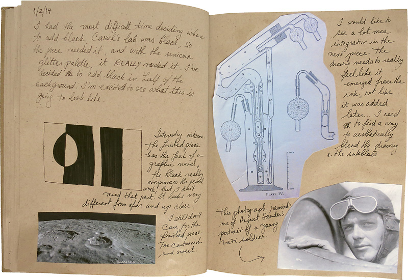
In the previous entry, I talked about my progress at the International Museum of Surgical Science, including an intuitive decision to cut ovals and rotate them 180 degrees. More recently, I realized that this cutting and repositioning is evocative of a surgical procedure, which is quite fitting for this residency. Each day, I find myself surrounded by artifacts used for slicing, clamping, stretching, and crushing. No wonder I had the urge to cut my inkblots!
After I spent a few weeks sketching various objects in the display cases, browsing through the storage space, looking at glass slides of eye surgery diagrams, and developing the oval cutout method, I finally decided to begin the first piece by focusing on one particular artifact in the museum collection: the Lindbergh perfusion pump.
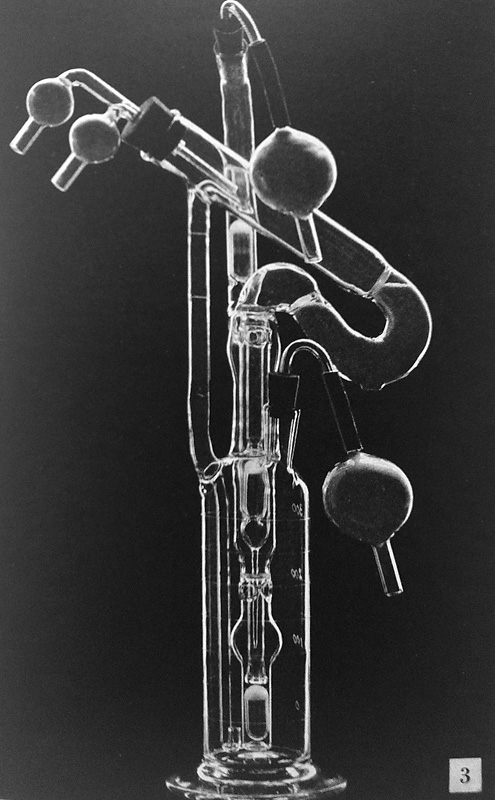
There were several reasons for my choice. First, I was unaware of this object’s intended use, which instantly drew me to it; secondly, I noticed that the curator, Lindsey Thieman, often expressed excitement about it while doing a walk-through of the collection; and finally, the object looked a lot like the imagined glassware that got me started on this body of work in the first place, back in 2002.
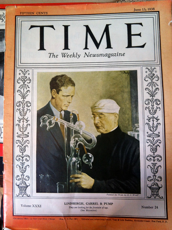
The Lindbergh perfusion pump turned out to be much more bizarre and uncanny than I expected it to be. I thought I would find out that the pump was used for an ordinary medical procedure involving blood vessels or the heart—since it was located in the vascular-themed room. Instead, I discovered that the pump was a collaboration between the famous aviator, Charles Lindbergh, and a French surgeon, Alexis Carrel, both of whom were interested in devising a method to keep organs alive outside of the body using a kind of artificial heart. Their book, The Culture of Organs, describes the procedure for keeping animal organs alive inside the pump, while the June 13th, 1938 issue of TIME magazine has a great description of the laboratory setting: “A secluded labyrinth of black, dustless, germless laboratories zigzags across the top floor of the main building of the Rockefeller Institute for Medical Research in Manhattan. Black are the floors, black the furniture, dark grey the windowless walls, shadowless the bleak illumination that comes through the skylights. Entrance to this aseptic, dustless, reflectionless hideaway is by a spiral staircase from an anteroom on the floor below. Only scientists particularly interested in fractioning life to its lowest common denominators may mount that spiral. And all must wash their hands and faces, put on gowns and hoods of black cloth – all except the master of this pure and dark domain, master of its purified and black-clad servants. He, the most famed member of the Rockefeller Institute, Dr. Alexis Carrel, distinguishes himself from the others with a little white headpiece that looks like the hat of a U.S. bluejacket.” If this wasn’t strange enough, in another article of September 16th, 1935, TIME magazine describes a “High Council of Doctors” proposed by Carrel “to which the political leaders of the world would come for their orders.”
Despite the strange and horrifying stories, I found it especially interesting that the reason why Lindbergh joined Carrel in search of an artificial heart was Elisabeth Morrow, Lindbergh’s sister-in-law, who had a heart condition before open-heart surgery was feasible. What struck me among all the madcap theories and grand plans was the simple ordinary story that centered on one individual’s medical condition that set in motion the search for an artificial heart.
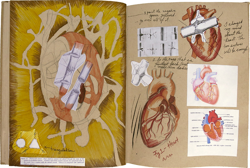
After I finished my first drawing/painting, out of sheer curiosity I continued reading about Carrel, and discovered that his reason for going into vascular suturing—and ultimately winning a Nobel Prize for it—was yet another individual’s medical condition. After Carrel found out that the popular French president in the late 1800’s, Sadi Carnot, died due to a stab wound that severed large irreparable veins in his abdomen, Carrel made it his life’s work to figure out how to suture these types of blood vessels seamlessly. He devised something called the triangulation method, in which the edge of the severed blood vessel is stretched out in three directions in order to properly sew the ends together. This method is allegedly still in use today.
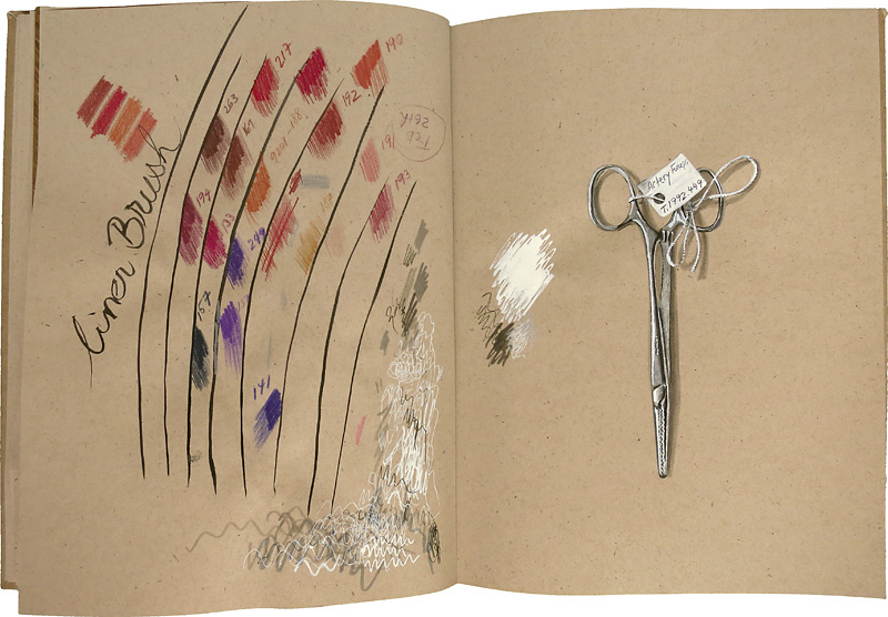
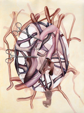
My second piece at the museum centered on this story. In order to add an image of artery forceps to this piece, I observed a physical pair of forceps in the museum collection and rendered it in metallic silver colored pencil.
I titled each work by the name of the individual who set the desire for medical invention in motion: Elisabeth Morrow, and Sadi Carnot. The implicit bodies of these individuals are depicted in a quasi-medical-illustration style, while the oval cutouts located the depictions in the context of a portrait.
In search for a title for the series, I began with a simple description of the work: all of the work begins with an oval cutout, and represents a portrait. An internet search with these key words immediately pulled up The Oval Portrait by Edgar Allan Poe, which was quite a surprise to me. What shocked me even more was that this short story was about art, representation, and subjectivity. Less shocking were the topics of death and doom; it is, after all, Poe.
What a fascinating post. I learned so much from it. It’s funny you should mention “The Oval Portrait” since I just covered it with an ACT student I was tutoring.
How fantastic! I’ve always enjoyed the way your art overlaps with anatomy and science, and I really love this blending with history and archival. I also think the personalizing with those whose bodies inspired the tools lends a humanity to it.